| Japanese | English |
Auscultation of a breast is very common. Organs which make sound in a breast are two, lungs and the heart. Other illnesses may be able to be diagnosed. The sound of lungs and the heart is very complicated. So the qualities of diagnosis are very different between an experienced doctor and an inexperienced doctor. The relation between lung disease and lung sound is summarized here.
1. Pneumonia
This is the state in which bacteria and the virus were infected and bred in
lungs. From the state of respiratory sound, it can be diagnosed about the grade
and cause bacillus of pneumonia. The feature of sound is coarse crackle, which
was analyzed in Lung sound measurement 2. More precisely, it is heard by
bronchitis, pneumonia, pulmonary tuberculosis, pulmonary infarction, pulmonary
suppuration, a lung congestion, the lung blister, bronchiectasis. Crackle sound
is described as fine, popping, crackling, discontinuous, non-musical noises. It
lasts about 10-25 ms.
2. Emphysema
Emphysema is the disease in which respiratory bronchioles and alveoli extend and
are destroyed. Consequently, effective lung capacity falls and the respiratory
function becomes bad. Emphysema is caused by smoking addictions and is
increasing in recent years.
3. Bronchial asthma
A characteristic sound can be heard. So it can be found easily. Auscultation may
be the most effective diagnostic method. The feature of sound is wheezing.
Wheezes are caused by narrowing, constriction, or spasm in the very small
airways. They can occur because of asthma, congestive heart failure, fibrosis,
pneumonia, and tuberculosis, also. The lasting time of the wheezing sound is
about 250 ms.
4. Pneumothorax
This is a disease in which a hole opens suddenly in lungs, air leaks in the
thorax, and lungs are pressed and shrunk. It can be found immediately since
respiratory sound stops. When time is taken by the X-ray examination, dyspnea
may advance suddenly and may cause shock condition. This is the illness which
should be diagnosed only by auscultation.
5. Pleurisy
Pleural membrane is a film covered with lungs. Pleurisy is the state in which
this film is infected with bacteria and inflammation arises. This can be
diagnosed by auscultation.
This time, lung sound is measured by a handmade stethoscope microphone as same as the heartbeat measurement. To secure a high S/N ratio, the stethoscope microphone was stuck on the body with the gummed tape.
| Date | 9 Apr. 2003 |
| Place | Home |
| Subject | 50 years old man |
| Microphone | SONY ECM-T150 Electric Condenser Microphone (Dimension: Diameter 6mm / Length 12mm) |
| Amplifier | SONY DAT WALKMAN TCD-D100 |
| PC | DELL INSPIRON 5000e |
| OS | Windows 2000 Professional |
| Software | DSSF3 |
| Other equipment | Stethoscope |
| Setup |  |
| WAVE file |
This is the peak level monitor at the time of no signal.
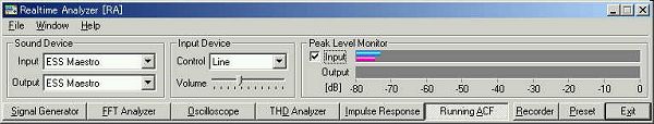
This is the background noise level measured with all equipments working and no input signal. It seems that the stethoscope microphone has high sensitivity. The level is between -28 and -35 dB.

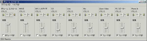
This is the Windows volume control.
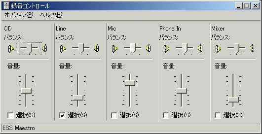
This is the input volume control. Line is checked.
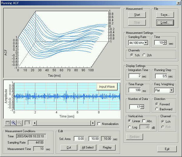
This is the running ACF measurement window. Click the Start button to measure the input signal from the microphone.
Then click the Save button. The "Save Measurement Data" dialog appears.
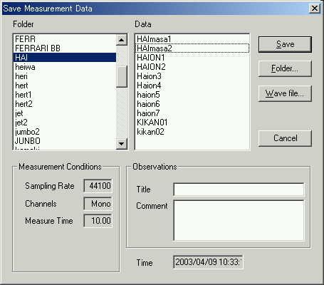
Measured data can be saved as the .wav format. The saved wave file here is a monaural data with 44.1 kHz sample rate.
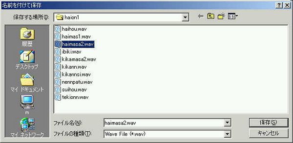
Wave file can be loaded from the running ACF window. Click the "Load" button and select the "Wave file". Sample data used in this page can be downloaded from here. Right click the link and save in the arbitrary directory.
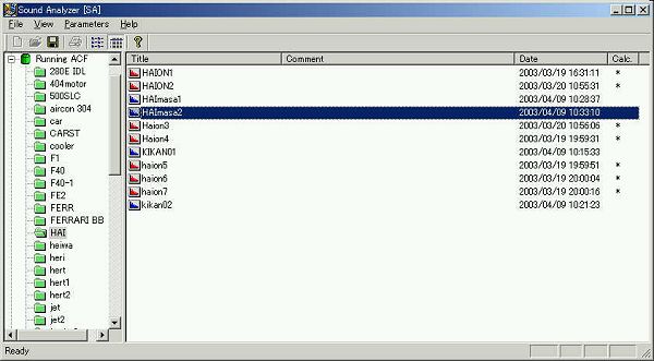
Saved data is loaded by SA.
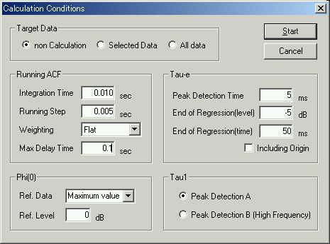
This is the calculation conditions window. Conditions are set for the high temporal resolution analysis.
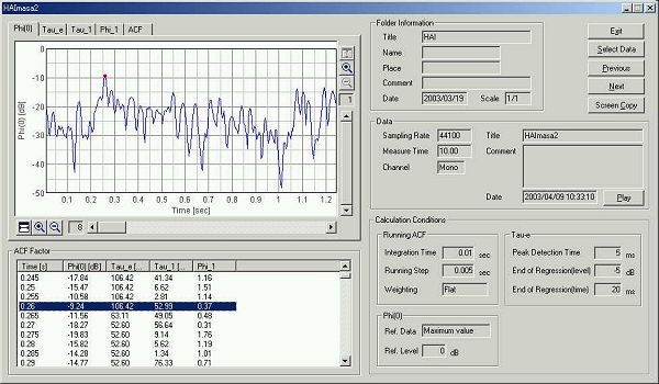
This is the analysis result. The graph Phi(0) shows the time course of the sound level.

The graph above is zoomed in at the first 1 second. The lung sound is repeated like this waveform. Heartbeat may also be included. I will not continue the analysis here.
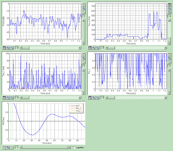
Time course of the acoustical parameters calculated from the ACF are shown below. This graph image is created by clicking the "Screen Copy" button.
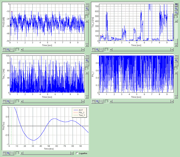
This is the time course of the ACF parameters for 10 seconds.
April 2003 by Masatsugu Sakurai